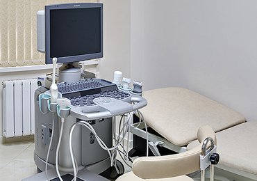
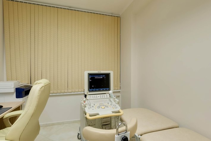
Equipment
Ultrasonic Test Department is equipped with the most advanced expert class devices from the leading manufacturers, such as Siemens-Acuson, Toshiba, Philips.
Scanners used in our department has a fundamentally new structure of high-density beam formation, which guarantees the highest resolution and details of clinical images and a fast and reliable diagnostic result. The devices has a large set of powerful clinical tools that provide the improved quality of images, quantitative analysis and interventional procedures during clinical and scientific studies. The modular structure and ability to process raw data simplify upgrading and allow for introduction of the necessary adjustments at any time.
Technologies
-Volume ultrasound visualization is required both by physicians and patients. 3D/4D mode allows to display anatomical structures as a three-dimensional image in a natural way, which is easy for spatial perception. The new tool ensures the creation of a 3D image of exceptional quality with a high frame rate for an uncompromising workflow and clinical result. Data saved in the original format can be downloaded, viewed and processed later without affecting the study results. The technology ensures clear detailing and improved visualization of structures and cavities. 3D colour Doppler mode is not inferior to 2D mode by quality. 3D cavity inspection mode allows to invert a grey scale image to reproduce hollow structures in the form of surface reconstruction.
-Multiplanar reconstruction (MPR) — this function allows to visualize objects in three orthogonal planes simultaneously, together with the image of surface or volume reconstruction. This ensures a better understanding of the anatomical correlation of structures or assess the spread of pathological changes.
-Ultrasound tomography allows to get a series of parallel sections in any plane of a given volume in a moment. This is a very effective tool for assessing pathological foci and adjacent structures.
-SMI technology (expert microvascular imaging) enables to visualize microvascular structures with the slow blood flow, using only ultrasound. Previously, medical specialists were limited to the use of contrast agents or invasive methods for visualization of blood flow around tumor masses, lymph nodes, etc.
-Panoramic scanning and colour panoramic scanning. Technology of displaying a full image of a 3D object. This technology stipulates that the ultrasonic sensor is passed with a constant speed along the surface of the object under measurement, and the whole object is displayed on the screen.
-MicroPure — the function allows to detect microcalcifications, potential markers of malignant tumors of mammary glands and other organs. This method allows for automatic detection of microcalcifications in the form of white spots on darkened 2D images.
-Shear wave elastography — displays the tissue's mechanical properties, the difference in stiffness, elasticity and extensibility of healthy and atypical tissues. The technology is used to study formations of mammary glands, lymph nodes, thyroid gland, prostate gland, liver for early acquisition of data on the malignancy of pathological structures and deciding on objective feasibility of fine-needle aspiration biopsy, as well as determination of the stage of fibrous changes of parenchyma.
-ARFI technology — ultrasound imaging using acoustic energy to determine the presence of volume formations and intensity of structural diffuse changes in the liver, for example, upon hepatic fibrosis or cirrhosis;
-External workstation. Supports the full access to clinical data and diagnostic tools at the right time and place. The integrated set of functions for raw data handling and a clinical programme package enables fast and easy viewing, analysis and archiving of data and creation of reports.
Unique basic technologies ensure the unsurpassed level of clinical accuracy, performance and usability. Diagnosis establishment is faster and more accurate now than ever before.
Scope of application
The department applies more than 70 diagnostic methods in such areas as:
angiology;
cardiology;
gastroenterology;
obstetrics and gynecology;
neurology;
traumatology and orthopedics;
urology;
oncology;
surface structures and organs;
transcranial studies;
Staff
Our department is staffed with 11 physicians who have a large professional experience. 4 physician have High-Level Certificate, 3 physicians — Category 1. 5 people are Candidates of Medical Science.
-
 on Lomonosovskiy
 -
 on Lomonosovskiy
Grigoryeva
Yevgenia OlegovnaUltrasound test physician
candidate of medical science
Length of service: 19 years
-
 on Lomonosovskiy
 -
 on Lomonosovskiy
Dubrovin
Yevgeny EduardovichHigh-Level Certificate
Ultrasound Test PhysicianLength of service: more than 20 years
-
 on Lomonosovskiy
Mutaev
Shakhmar MagomedovichHigh-Level Certificate
Ultrasound Test Physiciancandidate of medical science
Length of service: more than 20 years
-
 on Lomonosovskiy
Prelatova
Yuliya VadimovnaHigh-Level Certificate
Ultrasound Test Physiciancandidate of medical science
Length of service: more than 20 years
-
 on Lomonosovskiy
Simonova
Anna BronislavovnaHigh-Level Certificate
Ultrasound Test PhysicianLength of service: more than 20 years
-
 on Lomonosovskiy
 -
 on Lomonosovskiy
Yakovleva
Maria SergeevnaUltrasound Test Physician
Category 1candidate of medical science
Length of service: 20 years