Functional diagnostics is an area of medicine dealing with the assessment of functional capabilities and non-invasive diagnostics of affected organs and systems. Functional methods of study allow to detect diseases at both clinical and preclinical stages (when the disease does not cause troubles to a patient). Early diagnostics allows to prevent disease progression and to carry out the most effective treatment. Methods used in functional diagnostics are informative, safe and can be repeatedly performed for different categories of patients.
Main tasks of the functional diagnostics room:
- examination of practically healthy persons to determine the functional state of the body;
- - examination of patients with risk factors and detection of pathology at early preclinical stages of the disease;
- - assessment of functional and anatomical changes in organs and systems at different stages of the disease;
- - performance of functional, pharmacological and stress tests to select adequate therapy;
- - analysis of effectiveness of therapeutic measures.
Methods of study conducted in the functional diagnostics room.
Cardiovascular studies:
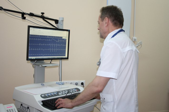
Electrocardiography (ECG) at rest.
ECG recording of the electrical cardiac activity allows to assess the functional state of the myocardium, is used for differential diagnostics of various types of arrhythmia, coronary artery disease, myocardial infarction, stenocardia and other cardiac diseases. Electrocardiogram recording is carried out both in the ECG room and in the wards using portable electrocardiographs. The advanced Schiller multichannel electrocardiographs (Switzerland) are used for ECG recording.
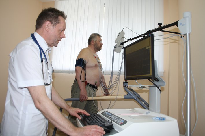
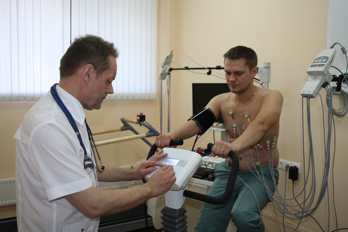
Graduated exercise tests: treadmill stress test, bicycle ergometry, stress ECG tests.
Testing is the most reliable non-invasive method of detection of latent myocardial ischemia, exercise tolerance, blood pressure response to stress, which allows to detect the disease, select adequate therapy for ischemic disease and arterial hypertension, develop a rehabilitation plan. In accordance with the computer programme, the patient performs an increasing stress test based on the track speed and slope (walk uphill simulation). During the stress test and the recovery period, the continuous monitoring of the patient’s condition and hemodynamic parameters (continuous ECG monitoring, heart rate and blood pressure monitoring) is carried out. The study is conducted using a stress system with Series 2100 GE Healthcare treadmill (USA).
Transthoracic echocardiography (Echo-CG, cardiac ultrasound) with Doppler colour mapping, tissue Doppler imaging.
The method is based on ultrasound imaging of cardiovascular anatomical structures. Echo-CG allows to assess the anatomical and functional state of the cardiac muscle, valve apparatus, cardiac cavities and major vessels, to recognize congenital and acquired defects, assess global and local myocardial contractility, systolic and diastolic disorders, identify myocardial hypertrophy, intracardiac thrombosis, calcinosis, cardiac tumors, presence of fluid in the pericardial cavity. Echo-CG studies are conducted using an expert-class GE LOGIQ E9 ultrasound scanner (USA).
Nervous system studies:
Electroencephalography (EEG) - method of recording of brain biopotentials, assessment of the electrical activity of the brain upon its various states and diseases: fainting, loss of consciousness, to exclude or confirm epilepsy and monitor the effectiveness of its treatment, vascular, degenerative, inflammatory diseases, injuries, etc. EEG is a safe and highly sensitive method of assessment of the functional state of the brain. A 16-channel electroencephalograph is used for EEG studies.
Respiratory system studies:
Spirometry (PFT) - pulmonary function test using the flow-volume loop method, drug tests with bronchodilators.
-
 Voronovo Sanatorium